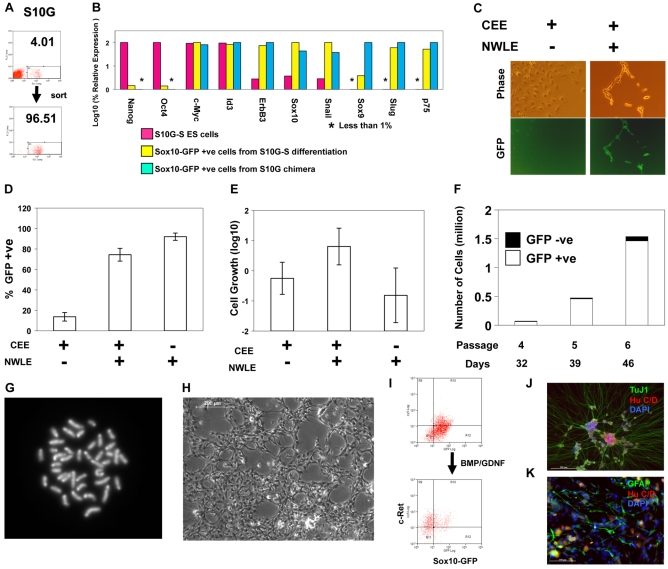
Fig. 3.
Culture of embryo-derived neural crest cells in the presence of noggin, Wnt3a, Lif and Et-3. (A) Flow cytometric purification of neural crest cells from E10.5 S10G chimaera. (B) qRT-PCR analysis of marker mRNA expression in: S10G-S ES cells; Sox10-GFP-positive cells purified from S10G-S embryoid bodies and cultured in NWLE for 5 days; Sox10-GFP-positive cells purified from E10.5 S10G chimaeras and cultured in NWLE for 7 days. The highest level of expression of each gene was defined as 100%. Bars represent means from duplicate PCR reactions. (C) Phase-contrast and fluorescence images of chimaera-derived neural crest cells in NCC medium with or without NWLE for 5 days. (D-K) Expansion of embryo-derived neural crest cells with blasticidin selection for Sox10 expression. (D,E) Proportion (D) and proliferative index (E) of Sox10-GFP-positive cells 6 days after purification from E10.5 S10G-B chimaeras. GFP-positive cells were detected by flow cytometry. Bars represent mean ± s.d. of six independent wells. Proliferation index was determined by dividing the number of GFP-positive cells by the initial cell number plated. (F) Long-term culture and growth of Sox10-GFP-positive neural crest cells purified from chimeric embryos and cultured in NWLE with blasticidin. Bars represent total number of cells at indicated days and passage number. The proportions of GFP-positive and -negative cells, as quantified by flow cytometry, are shown by white and black boxes, respectively. (G) Metaphase spread from neural crest progenitor culture in NWLE plus blasticidin. (H) Phase-contrast image of culture at passage 5 expanded for over one month. (I) Flow cytometry analysis of Sox10-GFP and c-Ret in cultured neural crest cells in NWLE and 5 days after transfer to N2B27 with Bmp and Gdnf. (J,K) Neuronal and glial differentiation of neural crest cells after 8 passages (over 50 days) in NWLE. Cells were transferred to medium containing bFgf/Bmp4/Gdnf for 5 days then fixed and immunostained for HuC/D, TuJ1 and Gfap. Scale bars: 200 μm in H,J; 100 μm in K.

