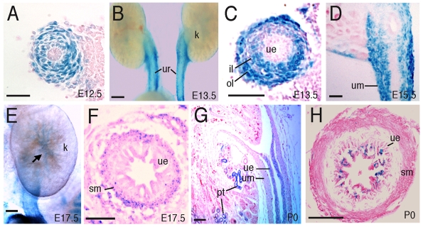
Fig. 1.
Expression of Six1 in the developing mouse ureter. Sections are X-gal stained for lacZ expression (blue). (A) A transverse section of E12.5 Six1+/lacZ ureter. (B) Whole-mount Six1+/lacZ kidney (k) and ureter (ur). (C) A transverse section of the ureter shown in B. (D) A longitudinal section of E15.5 Six1+/lacZ ureter. (E) Whole-mount E17.5 Six1+/lacZ kidney and ureter. (F) A transverse section of the ureter shown in E. (G) A longitudinal section of P0 Six1+/lacZ kidney and ureter. (H) A transverse section of the P0 Six1+/lacZ ureter. il, inner mesenchymal layer; ol, outer mesenchymal layer; pt, proximal tubules; sm, smooth muscle layer; ue, urothelium; um, ureteral mesenchyme. Scale bars: 50 μm.

