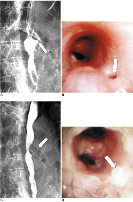
Fig. 1.
Esophagobronchial fistula due to pressure necrosis by esophageal stent.
Esophagogram (A) and bronchoscopy (B) after esophageal stent removal shows definite fistula (arrows) at proximal end of stent site.
Esophagogram (C) and bronchoscopy (D) after bronchial stent placement (arrows), shows successful closure of fistula.

