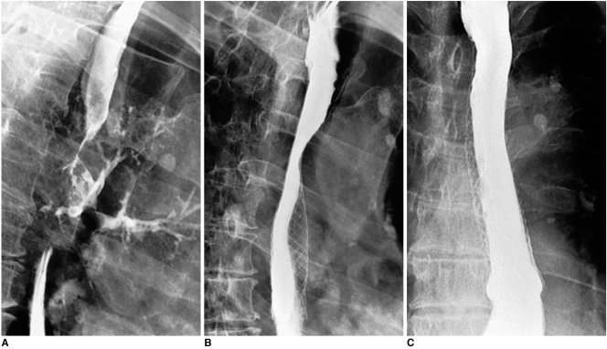
Fig. 2.
Esophageal cancer with development of esophagobronchial fistula. Ingested contrast medium is aspirated into left bronchi (A). Right anterior oblique (B) and anteroposterior (C) esophagograms obtained two days after placement of covered expandable metallic stent (18 mm in diameter), shows complete closure of fistula.

