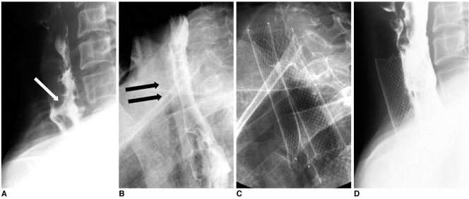
Fig. 4.
Esophageal cancer and esophagotracheal fistula.
A. Lateral esophagogram shows esophagotracheal fistula (arrow) and segmental luminal narrowing in cervical esophagus.
B. Radiograph obtained one week following esophageal stent placement shows diffuse tracheal narrowing (arrows).
C. Radiograph obtained following tracheal stent placement to relieve dyspnea.
D. Esophagogram obtained one week after tracheal stent placement shows good flow of contrast medium through esophageal stent without visualization of fistula and fully expanded tracheal stent.

