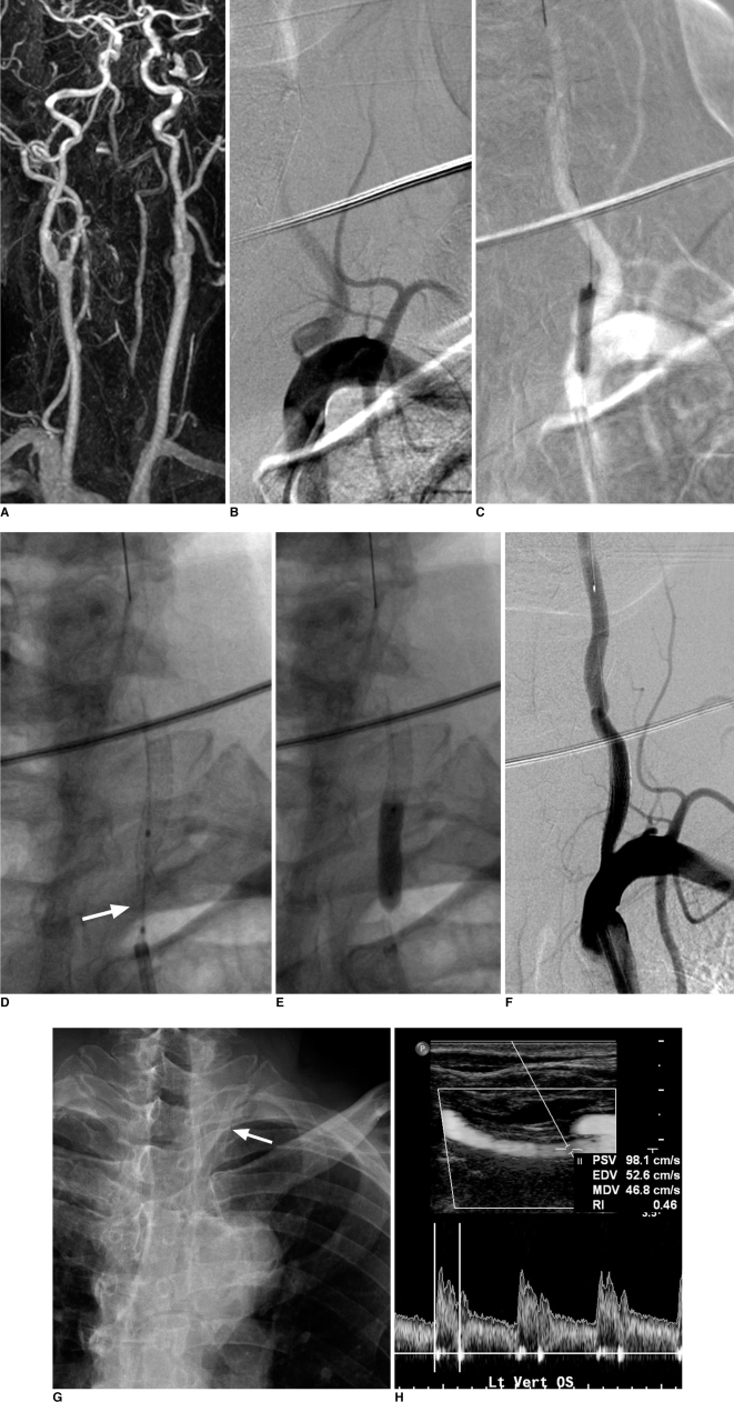
Fig. 1.
Placement of self-expanding stent in 68-year-old man who presented two weeks before procedure with right side weakness and ataxia due to multifocal embolic lesions in posterior circulation.
A. Contrast-enhanced MR angiography shows bilateral ostial stenoses. Left vertebral artery ostial stenosis lesion was considered to be cause.
B. Digital subtraction angiography shows focal severe stenosis of ostium.
C. Lesion is pre-dilated with 4-mm angioplasty balloon.
D. Self-expanding stent (Cordis Precise RX, 5 mm × 20 mm) is placed over stenotic lesion, which leaves residual stenosis (arrow).
E. Residual stenosis is successfully dilated using 5-mm balloon angioplasty catheter.
F. Final control angiogram shows minimal residual stenosis without flow restriction.
G. Chest radiograph obtained 12 months after procedure shows full expansion of whole stent.
H. Clinical and Doppler ultrasound follow-up performed 21 months after procedure shows good patency of stent without significant intimal hyperplasia.

