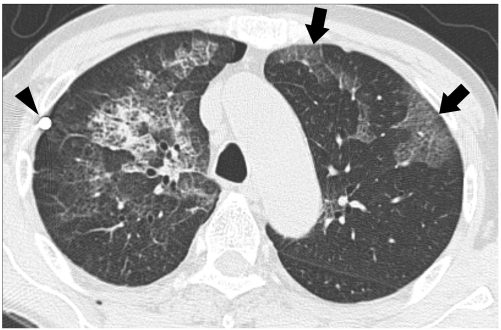
Fig. 3.
72-year-old man with bilateral re-expansion pulmonary edema. High-resolution CT taken seven hours after thoracentesis for right pneumothorax reveals mixed ground-glass opacity and minimal consolidation combined with intralobular reticulations and interlobular septal thickening; thus, suggesting ipsilateral reexpansion pulmonary edema. Like-natured opacities are also seen in contralateral lung (arrows), which consequently suggests contralateral re-expansion pulmonary edema. Note beam hardening artifact from tip of chest tube (arrowhead).

