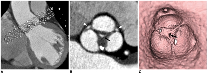
Fig. 2.
60-year-old male with mild central aortic regurgitation shown by transthoracic echocardiography.
A. Image shows batch reconstruction of aortic valve short-axial images in 1 mm thicknesses and 1 mm intervals.
B. Resultant image visualizes central coaptation failure zone of 0.17 cm2 (arrow) and fusion of left and noncoronary cusps. Small areas of coaptation failure in peripheral commissure of aortic valve were suspected (arrowheads).
C. Virtual angioscopic image confirms central regurgitation area (arrow). However, there was no evidence of commissural incompetency in periphery.

