Fig. 5.
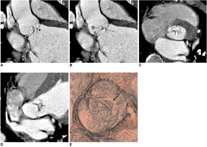
38-year-old male with right coronary cusp prolapse and eccentric aortic regurgitation.
A. Reformatted image shows prolapsed right coronary cusp (arrowhead) and coaptation failure of aortic valve (arrow).
B. Dotted lines A and B indicate image reconstruction planes for central aortic regurgitation and eccentric aortic regurgitation with prolapsed cusp, respectively.
C. Image reconstructed along plane A shows ovoid area (between arrows), indicating prolapsed part of right coronary cusp, not aortic regurgitant orifice.
D. Image reconstructed along plane B shows aortic regurgitant orifice (between arrows).
E. Virtual angioscopic image shows eccentric aortic regurgitant orifice (arrow).

