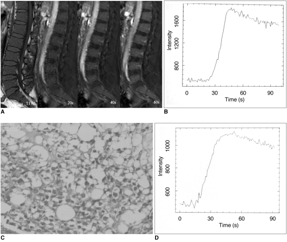
Fig. 2.
50-year-old patient with acute myeloid leukemia.
A. Pre-contrast T1-weighted spin-echo image of lumbar spine shows diffuse low signal intensity consistent with bone marrow infiltration. Dynamic contrast-enhanced MRI perfusion imaging of TFE-T1 weighted image obtained at 20 sec, 40 sec and 60 sec after gadopentetate dimeglumine bolus injection shows enhancement in vertebral bone marrow (Emax = 225.58%, ES = 10.74% and TTP = 22 sec).
B. L3 vertebral body of same patient shows TIC with rapidly rising slope (wash-in) during initial short period.
C. Bone marrow biopsy image of same patient (Fig. 3) with acute myeloid leukemia shows moderate tumor cell infiltration (Hematoxylin & Eosin staining, ×400).
D. Decreased Emax and ES values with increased TTP values (Emax = 135.35%, ES = 5.12% and TTP = 26.5 sec) were observed after treatment in same patient who responded well to treatment.
TFE = turbo field echo, Emax = peak enhancement percentage, ES = enhancement slope, TTP = time to peak, TIC = time-intensity curve

