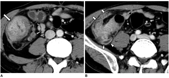
Fig. 10.
40-year-old man with cecal adenocarcinoma and he presented with acute pain in right lower quadrant and leukocytosis.
A, B. Contrast-enhanced axial CT scans show wall thickening with contrast enhancement in cecum (arrow) and thickened appendix (arrowheads). Surrounding fat stranding is severe (short arrows). Note pericolic enlarged lymph nodes (curved arrow). Cecal adenocarcinoma with invasion of appendix that resulted in acute appendicitis was pathologically confirmed.

