Fig. 12.
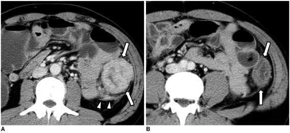
41-year-old man with adenocarcinoma of descending colon that was accompanied by distal segment of wall edema.
A. Contrast-enhanced axial CT scan shows large mass with contrast enhancement involving descending colon (arrows). Also note pericolic infiltration (arrowheads).
B. Axial CT scan obtained inferior to A shows mild annular wall thickening with preservation of wall layer in descending colon distal to tumor segment (arrows).

