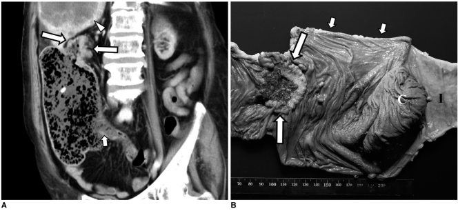
Fig. 2.
75-year-old woman with closed-loop obstruction secondary to adenocarcinoma of ascending colon and she had competent ileocecal valve.
A. Oblique coronal reformatted CT image shows obstructive mass in distal ascending colon (arrows) and marked dilatation of proximal colon that was filled with fecal material. Small bowel is not dilated and ileocecal valve area is indicated by short arrow. Also noted is hepatic metastasis (arrowhead).
B. Two days after CT scan, patient underwent emergency right hemicolectomy for her colon perforation. Photograph of resected specimen shows obstructive mass in ascending colon (arrows) and segmental dilatation of colon proximal to mass (short arrows). Perforation occurred just below colon cancer (not shown). C = cecum, I = terminal ileum.

