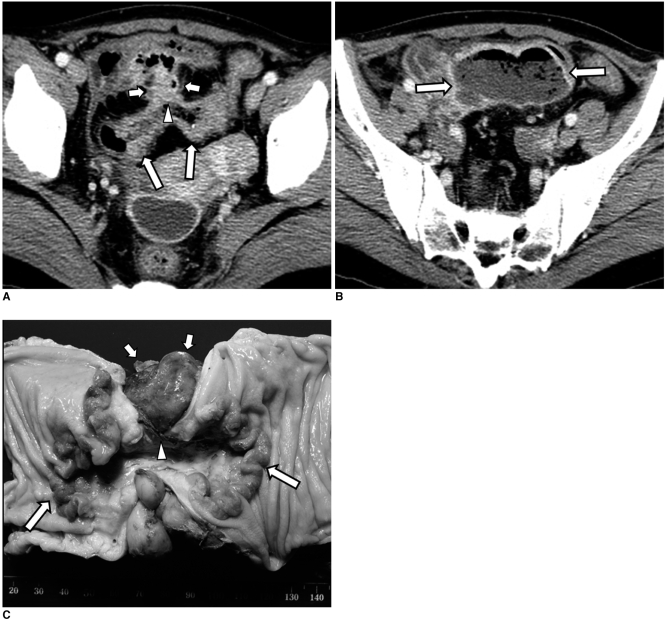
Fig. 3.
53-year-old woman with adenocarcinoma of sigmoid colon.
A, B. Contrast-enhanced axial CT scans show segmental wall thickening with contrast enhancement involving sigmoid colon (arrows in A). Anterior colon wall defect (arrowhead) with pericolic enhancing mass (short arrows) is identified. There is large abscess (arrows in B) in cranial direction to enhancing mass.
C. Photograph of specimen reveals ulcerofungating mass (arrows) with focal perforation (arrowhead). Pericolic inflammatory mass is also seen (short arrows).

