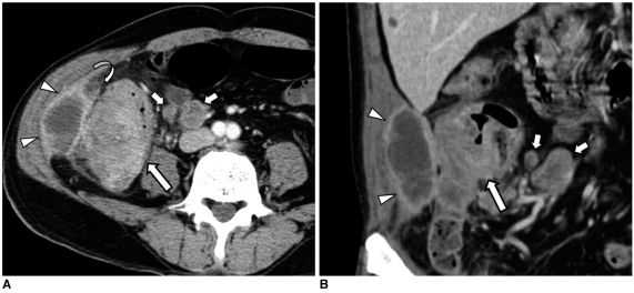
Fig. 5.
65-year-old man with adenocarcinoma of ascending colon.
A, B. Contrast-enhanced axial CT scan and coronal reformations show colon wall thickening with contrast enhancement (arrows), low-attenuated lesion of right paracolic gutter attached to abdominal wall (arrowheads) and adjacent fat stranding (curved arrow). Note pericolic enlarged lymph nodes (short arrows). Pericolic low-density lesion was surgically confirmed and it was pathologically diagnosed as inflammatory mass with abscess. There was no tumor involvement in peritoneal wall.

