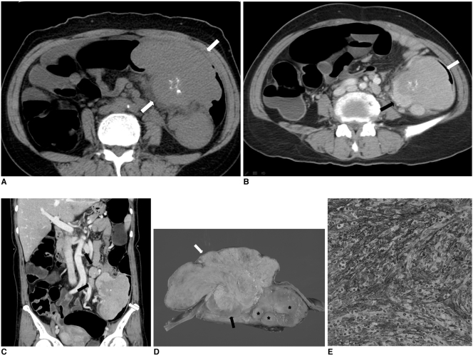
Fig. 2.
Follicular dendritic cell sarcoma in 52-year-old woman.
A. Pre-contrast axial CT image shows multiple foci of irregular and dense calcifications in mass (white arrows).
B. Post-contrast axial CT image shows well-defined large mass in mid-descending colon. Mass is composed of intraluminal (white arrow) and extraluminal (black arrow) components. Intraluminal component is less enhanced than extraluminal component.
C. Coronal reconstruction CT image shows several enlarged regional lymph nodes (black arrows) around mass (white arrow).
D. Gross specimen shows intraluminal component with relatively dull yellow color (white arrow) and non-necrotic area of extraluminal component (black arrow) with pink-tan color. Several enlarged pericolonic lymph nodes (asterisks) are noted.
E. Immunohistochemical staining for CD35 demonstrates positive cytoplasmic expression (×400).

