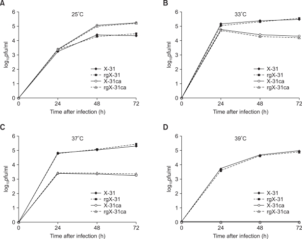
Figure 2.
Growth profiles of the X-31, rX-31, X-31ca and rX-31ca viruses in MDCK cells. MDCK cells were infected with 0.01 m.o.i. of the X-31, rgX-31, X-31ca and rgX-31ca viruses and incubated at 25℃ (A), 33℃ (B), 37℃ (C) and 39℃ (D) for 3 days. Supernatants were taken every 24 h for determination of viral titers by plaque assay.

