Figure 1.
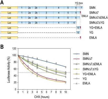
Delineation of YG + EMLA as a protein destabilization sequence in SMNΔ7. (A) Schematic diagram of Luc-fused SMN and a series of deletion constructs used for quantitative measurement of protein stability. Shown are SMN exon structures. YG box denotes the tyrosine/glycine (YG)-rich sequences in exon 6 of SMN. The EMLA sequence encoded by exon 8 is depicted by the red box at the C-terminal end of SMNΔ7. (B) 293T cells were transfected with plasmids expressing Luc-SMN, Luc-SMNΔ7, and the indicated deletion constructs. Forty-eight hours after transfection, the cells were treated with CHX (0.1 mg/mL) for various times as indicated, and then assayed for luciferase activity. Luc activity at each time point was calculated by comparison with those at time 0, which was set to 100%. Fifty percent activity is indicated by the gray dotted line. Error bars represent standard deviation (SD) from three independent experiments.

