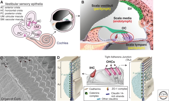
Figure 3.
Atypical tight-adherens junctions are found between outer hair cells and their supporting cells in the mammalian auditory organ (cochlea). (A) A cross section through the cochlea, which is the auditory organ, shows the three fluid-filled compartments, the scala vestibuli, scala media, and scala tympani. (B) Focus on the scala media that houses the auditory sensory epithelium, the organ of Corti (OC), which contains the mechanosensitive inner (IHC) and outer (OHCs) hair cells. sv, stria vascularis. (C) Top view of the organ of Corti, showing the highly organized hair bundles of IHCs and OHCs. (D) At the TAJ junction between OHCs and Deiters cell (DC), claudins partition into claudin 14- and claudin-9/6 containing subdomains that are distinguishable by their strand morphology and their associated cytoskeletal network. Note the colocalization of catenin complexes with claudin-containing domains (modified, with permission, from Nunes et al. 2006).

