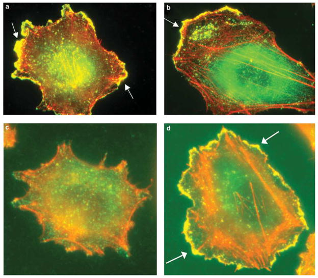
Figure 4.
Immunofluorescent staining with anti-cortactin Ab 4F11 (green) and F-actin (phalloidin, red) showed cortactin colocalization with F-actin in the advancing leading edges (yellow, arrows) in melanoma cell lines WM9 (a) and WM11158 (b). The same markers showed re-distribution of cortactin to the membrane ruffles (arrows), colocalizing with F-actin (yellow) in melanoma cell line 1205Lu after serum stimulation (d). Control serum-starved 1205Lu cell (c).

