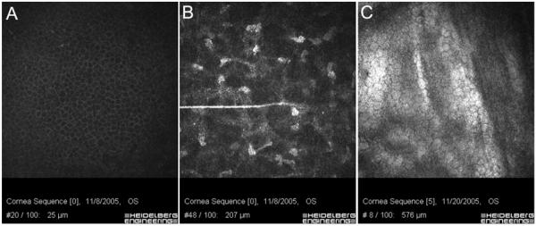
Figure 2.
HRT-RCM images of the wing cells (A), stromal cells (B) and endothelium (C) obtained by focusing in and out of the cornea using the remote-controlled lens drive. Since image acquisition was performed using the Heidelberg Eye Explorer software, the focal plane position, as detected by the inductive displacement transducer, is saved with each image (shown on bottom of each image). Image width = 400 μm.

