
Figure 1.
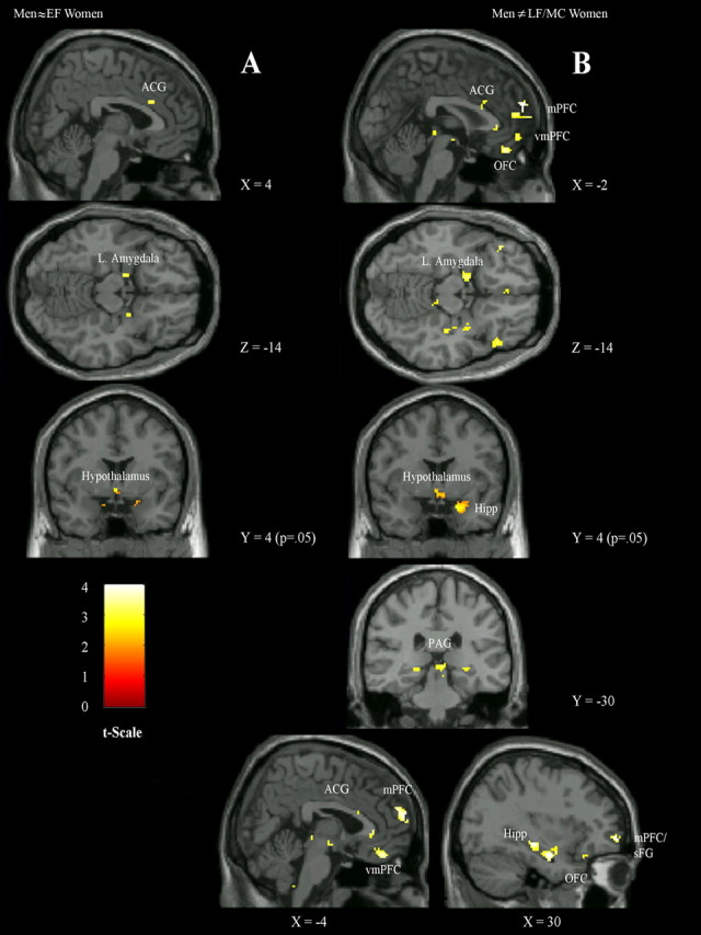
Significant sex differences in stress response regions comparing men versus women in early follicular (A) and late follicular/midcycle menstrual (B) phases. sFG, Superior frontal gyrus; Hipp, hippocampus; Hypothalamus, lateral hypothalamic area and ventromedial nucleus of the hypothalamus. Activations of hypothesized regions of interest were derived from 8 mm sphere around activation center; hypothalamic areas were from 4 mm spheres. Activations in Figure 1 are selected from Table 3, representative of hypothesized regions that were significantly different between men and EF women and men and LF/MC women (Table 3, columns 6, 7). For illustrative purposes alone, the peak activation in each activation cluster has an uncorrected p < 0.005, except for the hypothalamus at p < 0.05.