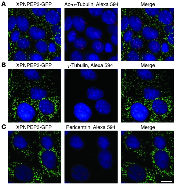
Figure 4. Full-length human XPNPEP3-GFP transiently expressed in IMCD3 cells localizes to mitochondria and not to the cilium/basal body/centrosome complex in IMCD3 cells.
Immunofluorescent microscopy was performed in IMCD3 cells that stably express human full-length XPNPEP3-GFP, which contains the mitochondrial signal sequence, demonstrating expression of XPNPEP3 in mitochondria. XPNPEP3-GFP was not detected in primary cilia (A), basal bodies (B), or centrosomes (C), when counterstaining with acetylated α-tubulin (Ac-α-tublin) (A), γ-tubulin (B), or pericentrin (C), respectively (middle and right panels). Scale bar: 10 μm.

