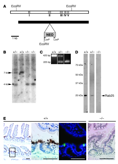
Figure 3. Construction of Rab25-knockout mouse.
(A) Schematic of the targeting construct for disruption of the Rab25 gene with insertion of the neomycin sequence into exon II of the Rab25 gene. The NEO cassette contained termination codons in all 3 frames. (B) Southern blot of EcoRV digests of DNA from heterozygote (+/–), homozygote wild-type (+/+), and 2 Rab25-knockout mice (–/–) probed with sequence from exon V. (C) 3 oligonucleotide PCR-based screening assays showed discrimination of Rab25-knockout mice from heterozygotes and wild-type littermate mice. (D) Western blot of extracts of protein (50 mg) from the gastric mucosa of littermate mice probed with rabbit anti-mouse Rab25 showing reduction of Rab25 expression in heterozygotes and complete absence of detectable Rab25 in Rab25-knockout mice. The distribution of molecular mass standards is indicated at left (kDa). The lane images shown are from noncontiguous lanes of the same gel and Western blot. (E) Immunohistochemical localization of Rab25 in the intestinal mucosa of wild-type mice (+/+) using either immunohistochemistry (left 2 panels) or immunofluorescence (third panel; green is Rab25 and blue is DAPI). The cells in the transition zone between the crypt and the villus stained most strongly for Rab25. In Rab25-deficient mice (–/–, far right), no Rab25 staining was observed. Scale bars: 50 μm.

