Figure 5.

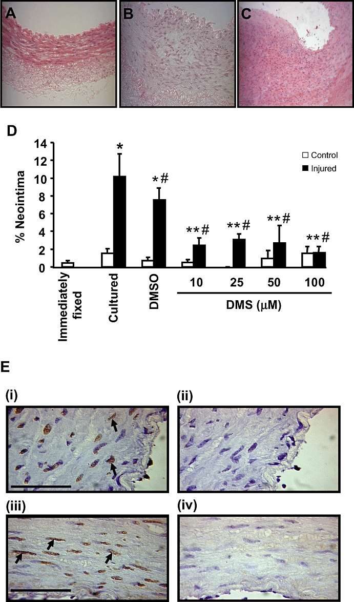
Effect of DMS on neointimal formation in vitro. Haematoxylin and eosin-stained sections showing the extent of neointimal formation in arterial segments that were (A) immediately fixed, (B) control cultured or (C) balloon injured and cultured. (D) Quantification of neointimal formation in balloon-injured LAD coronary arteries cultured in the absence and presence of DMS, 10–100 µM, n = 7–14). Values shown are mean + SEM, which *indicates P ≤ 0.05 compared to control, non injured cultured arteries using a Student's t-test; # indicates P ≤ 0.05 compared to injured cultured LAD coronary arteries using one-way anova followed by Tukey's test; ** indicates P ≤ 0.05 compared to injured, vehicle-treated cultured LAD coronary arteries using one-way anova followed by Tukey's test. (E) The effect of DMS on apoptosis. Apoptosis is marked by TUNEL staining (brown, marked with arrows) and counterstained (blue) with haematoxylin. Panels i and ii are arteries treated with 25 µM DMS. Panels iii and iv are artery sections which have been treated with 50 µM DMS. Panel i and iii have been treated with DNAse I to induce DNA strand breaks. All arterial sections received TUNEL staining. The extent of apoptosis with DNAse I + DMS was not different from DNAse I alone. Bar represents 50 µm.
