
Figure 1.
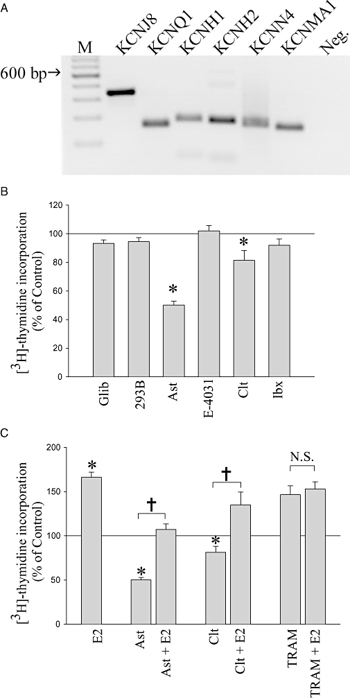
Detection of specific K+ channels expressed by MCF-7 cells and their contribution to basal and 17β-oestradiol (E2)-stimulated proliferation. (A) Agarose gel depicting PCR amplicons for the mRNA transcripts for KCNJ8 (336 bp), KCNQ1 (154 bp), KCNH1 (177 bp), KCNH2 (172 bp), KCNN4 (158 bp) and KCNMA1 (158 bp). Lane M is a DNA ladder, and Neg. is a negative control containing H2O instead of cDNA. (B) [3H]-thydimine incorporation by MCF-7 cells in the presence of K+ channel blockers specifically targeting these K+ channel types – glibenclamide (Glib, 30 µM), chromanol 293B (293B, 100 µM), astemizole (Ast, 3 µM), E-4031 (3 µM), clotrimazole (Clt, 10 µM) and iberiotoxin (Ibx, 100 nM). Mean normalized data from 3–11 experiments ± standard error of the mean. (C) [3H]-thymidine incorporation of MCF-7 cells in the presence of specific K+ channel blockers with or without co-application of E2. Concentrations of drugs were: E2 (1 nM), astemizole (Ast, 3 µM), clotrimazole (Clt, 10 µM) and TRAM-34 (TRAM, 10 µM). Mean normalized data from 4–28 experiments ± standard error of the mean. *P < 0.05 when compared with control, †P < 0.05, and N.S., not statistically significant between two indicated treatments.