
Figure 2.
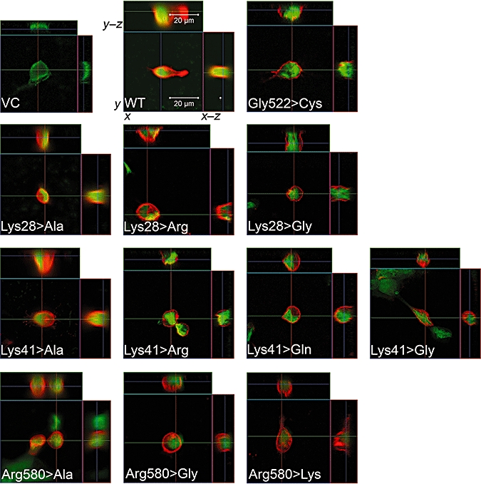
Immunofluorescence analysis by confocal microscopy of transiently transfected HEK293 cells expressing WT or mutant OATP1B3. VC: HEK293 cells transiently transfected with empty vector; WT: HEK293 cells transiently transfected with SLCO1B3 WT. The other pictures show the different mutants (e.g. Lys28>Gly, lysine at position 28 was mutated to glycine). All investigated mutants showed a clear localization of the protein in the plasma membrane. Only for the Arg580>Lys and the Arg580>Ala mutants that a partial retention in the cytoplasm was observed. For each mutant, the x–y, x–z and y–z dimensions are shown. Red fluorescence, localization of OATP1B3; green fluorescence, nuclei staining.