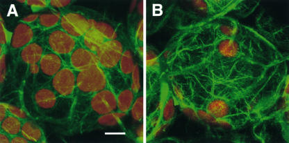
Figure 10.
Actin Cytoskeleton in Palisade Mesophyll Cells of the Wild Type and chup1-2.
The actin filaments were visualized in transgenic Arabidopsis plants by expressing GFP-mTalin with a confocal microscope in wild-type (A) and chup1-2 (B) backgrounds. The green and red signals show fluorescence from GFP and autofluorescence from chloroplasts, respectively. The images represent projections of 20 optical 0.95-μm sections. Bar = 10 μm.

