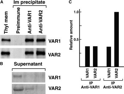
Figure 10.
Coimmunoprecipitation of VAR1 and VAR2.
(A) Detection of VAR1 and VAR2 in the immunoprecipitates. Thylakoid membranes solubilized by 0.8% DM were incubated with preimmune serum, Anti-VAR1, or Anti-VAR2. Each immunoprecipitate (Im precipitate) was subjected to immunoblot analysis with the antibodies indicated at right. Each lane for immunoprecipitate contained one-fifth of the total immunoprecipitate from thylakoid membranes, equivalent to 5 μg of chlorophyll. A sample of thylakoid membranes equivalent to 0.75 μg of chlorophyll was loaded simultaneously (Thyl mem). We repeated the experiments at least five times and obtained similar results. Results from a representative experiment are shown.
(B) Detection of VAR1 and VAR2 in the postimmunoprecipitate supernatants. Samples of the postimmunoprecipitate supernatants by Anti-VAR1 and Anti-VAR2 were probed with the antibodies indicated at right. The signals detected were very weak compared with those in (A). We found that a large amount of albumin from antisera comigrated with VAR1 and VAR2 on SDS-PAGE and prevented efficient blotting and immunodetection.
(C) Relative amounts of VAR1 and VAR2 immunoprecipitated by Anti-VAR1 and Anti-VAR2. The signals detected by immunoblot analysis in (A) were calibrated according to a series of dilutions of the VAR1 or VAR2 fusion protein loaded simultaneously (not shown). The relative amounts of VAR1 and VAR2 proteins included in the VAR1 or VAR2 immunoprecipitate (IP) are indicated.

