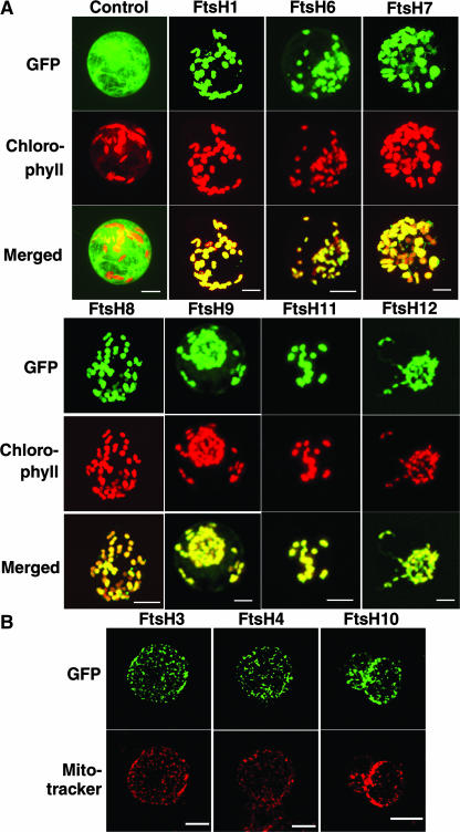
Figure 2.
Cellular Localization of the GFP Fusion Protein in Tobacco Protoplasts.
(A) SC tobacco protoplasts transformed with the GFP fusion. Genes used for the fusion constructs are indicated at top. Control indicates GFP with no targeting signal. GFP fluorescence, chlorophyll autofluorescence, and merged images are shown.
(B) SL tobacco protoplasts transformed with the GFP fusion. Genes used for the fusion constructs are indicated at top. Signals from GFP fluorescence and staining with MitoTracker are shown.

