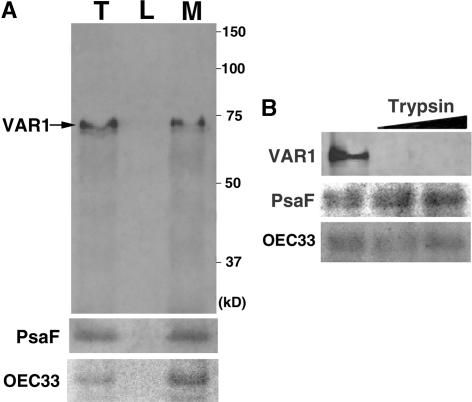
Figure 6.
Localization of VAR1 in Thylakoid Membranes and Its Topology.
(A) Immunoblot analysis of thylakoid proteins probed with Anti-VAR1 and control proteins (PsaF and OEC33). Purified thylakoids (lane T) were lysed and separated into soluble lumen (lane L) and insoluble membrane (lane M) fractions and separated by 10% SDS-PAGE. Proteins loaded in lanes L and M are derived from equal amounts of the thylakoid protein loaded in lane T. The band corresponding to VAR1 is indicated by an arrow. The positions of standard molecular marker proteins are indicated at right.
(B) VAR1 is sensitive to trypsin digestion. Purified thylakoids were incubated with two different concentrations of trypsin (slanted bar), separated by SDS-PAGE, and immunodetected by Anti-VAR1 and control antibodies against PsaF and OEC33.

