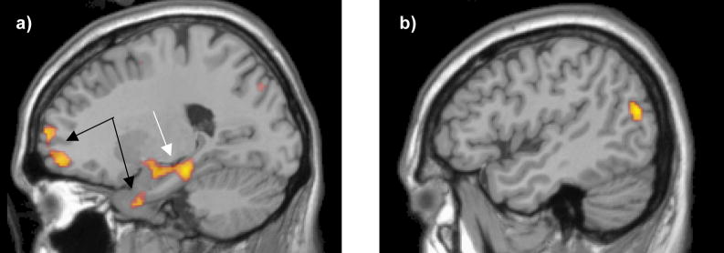
Figure 2. Areas yielding significantly greater activation for the SCP group than controls.
a) Slice shown at x = −21 (MNI space). Prefrontal regions and regions of the uncus denoted by the black arrows yielded a group differences because these regions were deactivated in controls but not patients. Parahippocampal and amygdalar regions denoted by the white arrow yielded a group difference because they were activated in patients b) Slice shown at x = −47 (MNI space). This lateral left temporal region yielded a group difference because of activation by patients but not controls.

