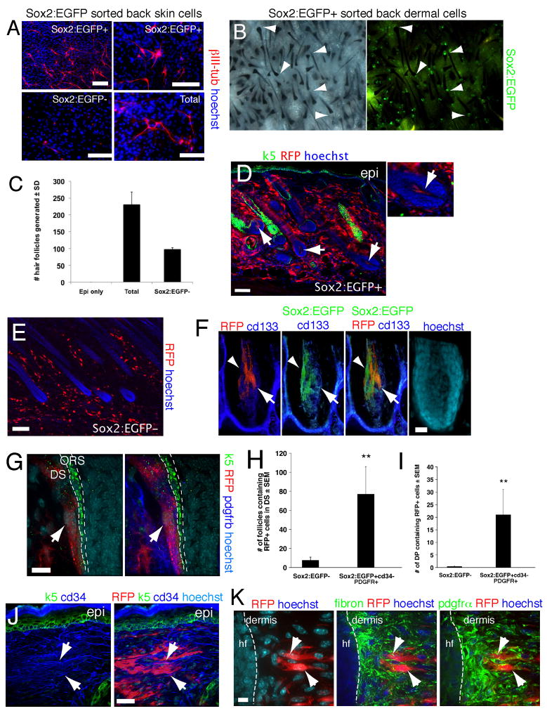
Figure 2. Sox2:EGFP prospectively identifies endogenous dermal precursors that induce hair follicles, home to a hair follicle niche, and differentiate into neural and dermal cell types.
(A) Sorted Sox2:EGFP+ and negative neonatal back skin cells differentiated for 12 days under neural conditions, and immunostained for III-tubulin (red). Total cells were sorted but gated only for live cells. (B) Patch assays using sorted Sox2:EGFP+, cd34− neonatal back skin dermal cells. The left panel shows brightfield and the right fluorescence illumination. Arrowheads denote EGFP+ DP. (C) Number of hair follicles generated in patch assays with total or Sox2:EGFP− neonatal dermal cells (n = 3). (D–K) Sorted, uncultured Sox2:EGFP+, cd34−, PDGFRα+ (D,F–K), or Sox2:EGFP− (E) neonatal back skin cells were transplanted into adult NOD/SCID mouse back skin, and analyzed after 2 weeks. All sorted cells were infected with an RFP-expressing retrovirus. (D) Immunostaining for RFP (red) and keratin 5 (green). Arrows denote follicle DP containing transplanted cells, with the right panel at higher magnification. (E) Immunostaining for RFP (red) on skin transplanted with Sox2:EGFP− cells. (F,G) High magnification confocal images of hair follicles immunostained for RFP (F,G, red) and Sox2:EGFP (F, green) plus the DP marker cd133 (F, blue), or keratin-5 (G, green) plus PDGFRβ (G, blue). In (F) the arrowhead and arrow denote Sox2:EGFP+ and cd133+ cells, respectively. In (G) the arrow denotes RFP+, PDGFRβ+ cells in the DS. ORS = outer root sheath. (H,I) Number of hair follicles (H) or follicle DP (I) containing RFP+ transplanted cells as shown in D–G. **P<0.01. (J,K) Immunostaining of interfollicular dermis for RFP (red) and cd34 (J, blue) plus keratin 5 (J, green) or fibronectin (K, pseudocolored green, center panel) plus PDGFRα (pseudocolored green, right panel). Arrows show cells positive for RFP and cd34 (J), or for RFP, fibronectin and PDGFRα (K). hf = hair follicle. Nuclei were stained with Hoechst 33258 (blue), as indicated. Scale bars = 100μm in A,D,E, 25μm in F,J and 10μm in G,K. See also Figure S2.

