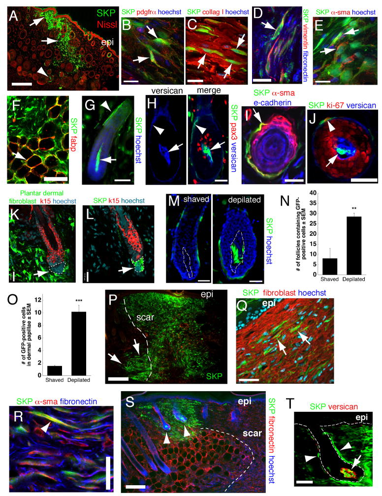
Figure 4. SKPs regenerate the dermis and integrate into a hair follicle niche upon transplantation into adult skin.
(A,B) Back skin transplanted with YFP-tagged neonatal mouse SKPs 2 weeks earlier and immunostained for YFP (green) and PDGFRα (B, red). Arrows and arrowheads in (A) show transplanted cells in the interfollicular dermis and the DP and DS of follicles, respectively. Arrows in (B) denote double-labeled cells. (C–E) GFP-expressing adult rat SKPs transplanted into depilated adult NOD/SCID mouse dermis 21 days earlier, and immunostained for GFP (green) and collagen type 1 (C, red), vimentin and fibronectin (D, red and blue, respectively), or α-sma (E, red). Arrows denote double-labeled (C,E) or triple-labeled (D) cells. (F) GFP-tagged cells (green) within the hypodermis expressing the adipocyte marker fatty acid binding protein (red, arrow). (G–J) Hair follicles containing neonatal murine YFP+ SKPs 2–4 weeks post-transplantation as in (A). (G) Hair follicle with YFP-labeled cells (green) in the DP (arrow) and DS (arrowhead). (H) Follicle triple-labeled for YFP (green), the DP marker versican (blue) and the melanoblast marker pax3 (red). The arrow and arrowhead indicate the DP and DS. (I,J) Cross-sections of follicles showing transplanted cells (green) in the DS (arrow in I, arrowhead in J) expressing -sma (I, red) or Ki67 (J, red), but not e-cadherin (I, blue, an epidermal marker). In (J), DP cells (arrow) are positive for versican (blue). (K,L) Telogen follicles in skin transplanted 4 weeks earlier with GFP-tagged rat plantar dermal fibroblasts (K) or SKPs (L), immunostained for GFP (green) and keratin 15 (red). Only SKPs are in the DP (arrows, denoted by hatched lines). (M) Representative hair follicles containing neonatal murine YFP+ SKPs (green) 3 weeks after skin was shaved or depilated. Hatched lines denote the DP. (N,O) Number of follicles containing GFP+ cells (N) or GFP+ cells within the DP of individual follicles (O) following transplantation of adult rat GFP+ SKPs into depilated (n=4) versus shaved (n=4) skin. **p<0.01, ***p<0.001. (P–T) Skin 3 (P–R) or 4 (S,T) weeks after transplantation of neonatal YFP-tagged murine SKPs (green in all panels) adjacent to a back skin punch wound. In (P), transplanted cells are present at the site of injection (arrows), and within the regenerated tissue (denoted by dashed lines). (Q,R) Transplanted cells immunostained for fibroblast-specific antigen (Q, red, arrows), or for α-sma and fibronectin (R, arrowhead, red and blue, respectively). (S,T) Transplanted cells are also present in peg-like hair follicles at the boundary of the wound (S, arrowheads) within the DP and DS (T, arrow and arrowheads). Cells in the DP express versican (T, red). Some sections were counterstained with Hoechst 33258 (blue) or fluorescent Nissl (red), as indicated. Epi = epidermis. Scale bars = 200μm in A,P,S, 16μm in B–E, 25 μm in G-M,Q, 50μm in F,R,T. See also Figure S4.

