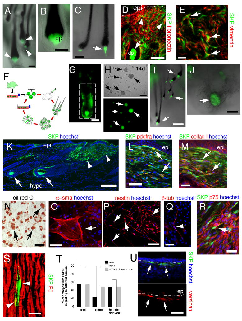
Figure 6. (A–E) Clonally-derived SKPs reconstitute the dermis and induce hair follicle formation.
Analysis of one adult rat GFP+ SKP clone in patch assays (A–C), or by transplantation into adult mouse dermis (D,E). After 2–4 (A,B) or 11 months (C) of culturing, clonal SKPs (green) induced the formation of hair follicles, where they comprised the DS and DP (arrowheads in A, arrow in C). (D,E) After 12 weeks in culture, cells from the same clone were transplanted and skin immunostained for GFP (green) and fibronectin (D, red, arrowheads) or vimentin (E, red, arrows). Arrow in (D) denotes the DP. (F–U) SKPs isolated from their hair follicle niche self-renew, serially reconstitute hair follicles and remain multipotent. (F) Schematic showing the serial reconstitution assay of hair morphogenesis. (G,H) A follicle isolated from a patch assay using adult rat GFP-labeled cells. Tagged cells isolated from the boxed area generated GFP+ SKP spheres (H, arrows) after 12 days in culture as seen by phase (top) and fluorescence (bottom) illumination. (I) Cells from these follicle-derived spheres induced formation of secondary hair follicles in the patch assay (arrows). (J) In tertiary follicle reconstitutions, follicle-derived SKPs generated hair follicles, but many also aggregated into DP-like structures surrounded by black melanocytes (arrow). (K–M) Skin transplanted with GFP+ follicle-derived SKPs for 4 weeks. Transplanted cells (green) integrated into follicle DS and DP (K, arrows) and contributed to the dermis (K, arrowheads), where they expressed PDGFRα (L, red) and collagen type 1 (M, red). Arrows indicate double-labeled cells. (N,O) Follicle-derived SKPs generated adipocytes, as indicated by the lipophilic dye oil red O (N, arrows), and sma+ cells, potentially myofibroblasts (O, arrow) in mesodermal differentiations. (P,Q) When differentiated under neurogenic conditions, they generated nestin+ cells after 5 days (P, red, arrows), and III-tubulin positive cells after 14 days (Q, red, arrow). (R,S) Sciatic nerve sections 6 weeks after transplant of follicle-derived SKPs, showing that some transplanted cells expressed the Schwann cell markers p75NTR (R, arrow) and P0 (S, arrowheads). (T,U) Transplantation of follicle-derived SKPs into the chick neural crest migratory stream (H.H. stage 18). (T) Quantification after 3 days in ovo showed follicle-derived (n=6) and clonal (n=8) SKPs behaved like total SKPs (n=9), migrating to the ventral nerve or DRG and skin, with some remaining close to the neural tube. (U) After 8 days in ovo, some of the transplanted cells that were in the dermis (green) were versican+ (red, arrows). Nuclei were stained with Hoechst 33258 (blue), as indicated. epi = epidermis. Scale bars = 100μm in A,B,C,D,H,P, 50μm in E,G,N,O,Q,U, 25μm in L,M,R,S, 200um in I,K, and 250μm in J.

