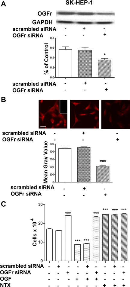
Fig. 4.
OGFr is required for OGF's inhibitory action on growth on HCC cells. A: Northern blot analysis and semiquantitative densitometry demonstrating the specificity and level of OGFr knockdown in SK-HEP-1 cells. Log phase cells were transfected for 24 h with either scrambled siRNA or OGFr siRNA. Forty-eight hours after transfection, cells were harvested and RNA isolated. Data (%OGFr/GAPDH ratio) represent means ± SE for 2 blots from independent experiments. *Significantly different from nontransfected cultures at P < 0.05. B: photomicrographs of log phase SK-HEP-1 cells stained with a polyclonal antibody to OGFr (B0344) demonstrating the extent of OGFr protein knockdown. Cells were transfected for 24 h with OGFr siRNA or scrambled siRNA, and incubated in media for an additional 48 h. Photomicrographs of cells stained with OGFr were taken at the same exposure time. Semiquantitative measurement of the OGFr immunohistochemistry demonstrating the level of protein knockdown in SK-HEP-1 cells. Decreased OGFr staining intensity (mean gray value) is indicative of decreased OGFr protein expression. Data represent means ± SE. ***Significantly different from nontransfected cells at P < 0.001. C: growth of SK-HEP-1 cultures transfected with OGFr siRNA or scrambled siRNA for 24 h and treated with either OGF (10−6 M), naltrexone (NTX; 10−6 M), or an equivalent volume of sterile water for 72 h; compounds and media were changed daily. Values represent means ± SE cell counts for at least 2 aliquots/well and least 2 wells/treatment. ***Significantly different at P < 0.001 from cultures that were not transfected and treated with sterile water.

