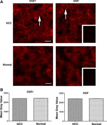
Fig. 7.
Presence and quantitative analysis of OGF and OGFr in surgical specimens of HCC and normal tissue. A: HCC resected from a 72-yr-old male with moderately differentiated cancer (Stage 3A) and stained with antibodies to OGF or OGFr. Note the immunoreactivity for OGF and OGFr adjacent to the nucleus and throughout the cytoplasm (arrows), as well as a speckling of immunoreactivity in the nucleus. Tissues processed with secondary antibody showed no staining (inset). Bar = 20 μm. B: semiquantitative densitometric analysis of OGF and OGFr from surgical specimens of 2 patients with HCC that compare tumor with normal (margin) tissues. Data represent means ± SE for at least 4 sections/specimen.

