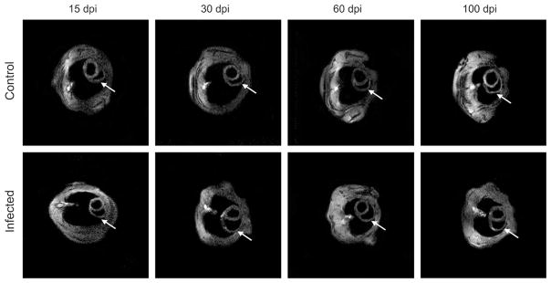
Figure 3.
Transverse magnectic resonance imaging (MRI) images of mice showing the short axis of the heart. There was no difference in left ventricular internal diameter during the period studied in infected mice compared with uninfected controls. However, the inner dimension of the right ventricle (RV) was significantly dilated from 30 to 100 dpi. The left ventricle wall thickness (LVWT) was increased at 100 dpi. There was no difference in LV wall thickness at 15, 30, and 60 dpi in comparison to uninfected controls. Arrows = RV.

