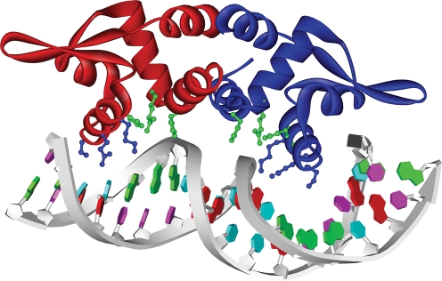
Fig. 5.
DNA binding: a model of PagR dimer binding to DNA with a bend of about 4 °. Mol.A is shown in red and Mol.B in blue. The side chains of the residues Lys64, Gln60 and Ser56 (blue) of the recognition helix point towards the major groove. The residues His26, Arg29 and His61 cluster together (green), indicating possible interactions with the DNA backbone.

