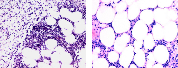
Figure 2.
Histology (painful node, left lower leg). Standard histological processing was performed on a painful node obtained from the lower limb. Two HE-stained sections at different magnifications (left 20×, right 30×) are depicted. The sections show predominantly lobular panniculitis with mixed inflammatory cell infiltration.

