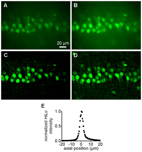
Figure 2. Optical sectioning with HiLo microscopy.
A–C. Speckle (A) and uniform (B) illumination images used in the calculation of the quasi-confocal HiLo image (C). D. z-projection of 12 HiLo sections (including C) separated by Δz = 2 µm. E. Measurement of the HiLo microscope axial resolution: integrated signal from a thin fluorescent layer (≈0.3 µm) as a function of defocus z. The measured axial resolution is 4 µm FWHM.

