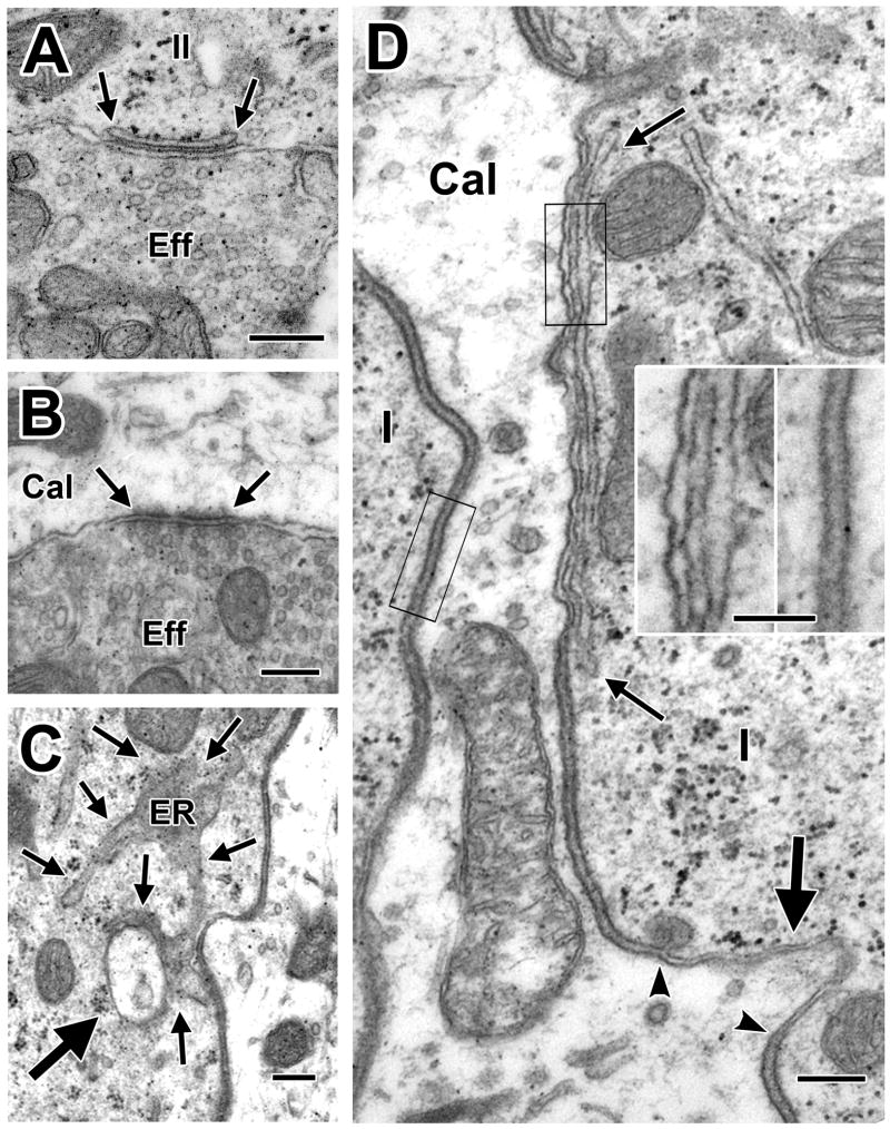
Figure 8.
Efferent innervation, invaginations and subsurface cisterns. A, B. Highly vesiculated efferent boutons contact a type II hair cell and a calyx ending, respectively. C. A calyx ending invaginates into a type I hair cell. The invagination, including a pinched off portion (thick arrow), is in close association with the endoplasmic reticulum (ER, thin arrows). D. At the location of a subsurface cistern (delimited by thin arrows) in a type I hair cell (I) adjacent to the inside wall of a complex calyx (Cal), there is a narrowing of the intercellular space, similar to that seen at a neighboring invagination (thick arrow, the narrow extracellular cleft of the invagination is delimited by arrowheads). The calyx membrane narrows roughly 60% near the cistern from about 8.5 nm, on average, to 5.2 nm, as seen in the insets taken from the areas indicated by the two black rectangles in D. Scale bars, 0.2 μm (A–D) and 0.1 μm (insets).

