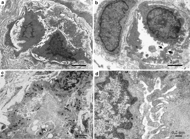
Fig. 2.
a Dermal capillary (case 2) showing discontinuous endothelial cells (ECs) displaying loose anchoring and formation of a second endothelial layer. b Dermal capillary (case 1) with an enlarged single EC. A prominently activated apical pole forms a transluminal bridge (arrows). Note the large pericyte on the left. c Dermal capillary (case 2) with the lumen almost completely obliterated by the enlarged, folded EC cytoplasm. Note the cytoplasmic transformation into a dense mesh and the numerous Weibel-Pallade granules. The thin basement membrane is almost invisible on the left side. d Dermal capillary (case 2) showing details of EC apical membrane folding together with numerous ribosomes and occasional microvesicles

