
Fig. 2.
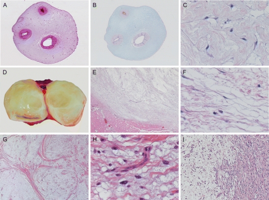
Characteristic macroscopy and histomorphology of the myxoid ECM. Rudolph Virchow introduced the term myxoma for those tumors morphologically resembling Wharton’s jelly of the umbilical cord (a), which contains large amounts of GAGs as detected by alcian blue (b). High-power image of Wharton’s jelly showing abundant myxoid ECM containing fibrillary collagens, interspersed between myofibroblast-like stroma cells (c). Intramuscular myxoma characteristically has a gelatinous appearance on cut surface (d) and is well circumscribed towards its peripheral tissue (e). On higher magnification, it shows the same abundant myxoid ECM as the umbilical cord (c) and no significant atypia of the sparse tumor cells (f). Histological criteria are still a hallmark of diagnosis, showing characteristic lobulated, hypocellular morphology of grade I myxofibrosarcoma at low magnification (g).Curvilinear blood vessels are often seen in grade I myxofibrosarcoma (but are not diagnostic), whereas tumor cells show vesicular, slightly atypical nuclei compared to intramuscular myxoma (h). Another hallmark of myxofibrosarcoma is areas with abrupt transition of grade (i) which was already mentioned by Mentzel et al. [95]