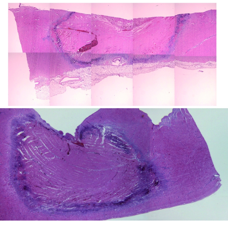
Figure 3.
Histological aspect of lesions in the right ventricle, as prepared for planimetric analysis. Haematoxylin eosin stain. Both sections show a large necrotic area, with an inflammatory margin, with a small central thrombus in the upper one. The upper lesion is the result of a magnetically steered application; the lower one from a standard catheter.

