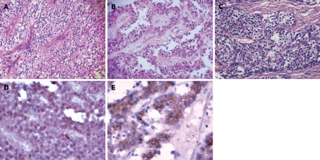
Figure 1.
Histopathological and immunohistochemical features of solid-pseudopapillary neoplasm (SPN) and pancreatic endocrine tumor (PET). A: SPN arranged in solid areas, patternless sheets of uniform epithelial cells with numerous small blood vessels (hematoxylin-eosin, original magnification × 200); B: SPN formed characteristic pseudopapillary changes due to the degenerative and discohesive nature of the tumor cells (hematoxylin-eosin, original magnification × 200); C: PET arranged in acinar-like pattern; the tumor cells are small and round with granular eosinophilic or clear cytoplasm (hematoxylin-eosin, original magnification × 200); D: β-catenin immunostaining of SPN: Nuclear translocation and accumulation of β-catenin protein (arrow) is seen in neoplastic epithelial cells (original magnification × 200); E: β-catenin immunostaining of PET: Membrane and cytoplasmic positive expression of β-catenin protein without nuclear stain (arrow) (original magnification × 200).

