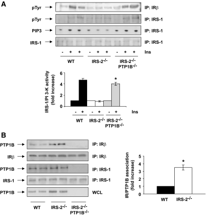
FIG. 5.
The lack of PTP1B in IRS2−/− mice restored IRS1-mediated PI 3-kinase signaling by releasing an IR/PTP1B association. A: Wild-type (WT), IRS2−/−, and IRS2−/−/PTP1B−/− mice (n = 9 animals per genotype) were fasted for 4 h, injected intraperitoneally with PBS or human regular insulin (Ins) (0.75 units/kg), and killed 15 min later. Livers were removed, and total protein extracts were prepared. Then, 600 μg total protein was immunoprecipitated with anti-IRβ and analyzed by Western blot with anti-pTyr. Total protein (600 μg) was immunoprecipitated with anti-IRS1 antibody and analyzed by Western blot with anti-pTyr and anti–total IRS1 or used for an in vitro PI 3-kinase assay. The conversion of PI to PIP3 in the presence of [γ32-P] ATP was analyzed by TLC. A representative experiment is shown. The autoradiograms showing PI 3-kinase activity were quantitated by scanning densitometry. Results are expressed as fold increase of PI 3-kinase activity and are means ± SEM. *P < 0.05, IRS2−/−/PTP1B−/− vs. IRS2−/−. B: Liver extracts from wild-type, IRS2−/−, and IRS2−/−/PTP1B−/− mice (n = 6 animals per genotype) were prepared and 600 μg total protein were immunoprecipitated with anti-IRβ or anti-IRS1 antibodies and analyzed by Western blot with the anti-PTP1B, anti-IRβ, and anti-IRS1 antibodies. Total protein (50 μg) was analyzed in whole cell lysates by Western blot with the antibody against PTP1B. The autoradiograms showing IR/PTP1B association from all animals analyzed were quantitated by scanning densitometry. Results are expressed as fold increase of IR/PTP1B association and are means ± SEM. *P < 0.05, IRS2−/− vs. wild type.

