FIG. 6.
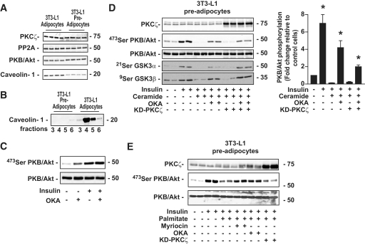
Effect of ceramide and palmitate on insulin-induced phosphorylation of PKB/Akt in 3T3-L1 preadipocytes. A: 3T3-L1 preadipocyte lysates were immunoblotted with antibodies against either PKCζ, PP2A, native PKB/Akt, or caveolin-1. B: CEMs prepared from 3T3-L1 preadipocytes were isolated as described in research design and methods. Equal amounts of protein (1 μg) were then immunoblotted with the antibody against caveolin-1. C: Preadipocytes were treated with 500 μmol/l OKA for 30 min and 100 nmol/l insulin for the last 10 min before being lysed. Cell lysates were immunoblotted with antibodies against either native PKB/Akt or Ser473 PKB/Akt. D: Control 3T3-L1 preadipocytes and KD-PKCζ–infected preadipocytes were preincubated with 100 μmol/l C2-ceramide for 2 h, followed by 500 μmol/l OKA for the last 30 min. Then, 100 nmol/l insulin was added to the cells for 10 min before being lysed. Cell lysates were immunoblotted with antibodies against either native PKB/Akt, Ser473 PKB/Akt, Ser21/9 GSK3α/β, and PKCζ. Scanning densitometry was performed to quantify changes in Ser473 PKB/Akt abundance in cell lysates. Bars represent mean ± SEM. *Significant change P < 0.05 relative to the untreated control. Blots shown represent three separate experiments. E: Control 3T3-L1 preadipocytes and KD-PKCζ–infected preadipocytes were preincubated with 0.75 mmol/l palmitate (conjugated with 0.2% [wt/vol] BSA) for 20 h. In some experiments, fatty acid incubation was also performed in the presence of 10 μmol/l myriocin. OKA (500 μmol/l) was added for the last 30 min and 100 nmol/l insulin for 10 min before being lysed. Cell lysates were immunoblotted with antibodies against either native PKB/Akt, Ser473 PKB/Akt, or PKCζ.

