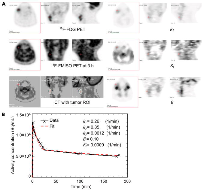
FIGURE 4.
Concordance between parametric Ki values suspected to exhibit no or low level of tumor hypoxia and low T/B in late-time image for patient 9. Transverse, coronal, and sagittal views are shown for 18F-FDG PET, 18F-FMISO PET at 3 h, CT fused with tumor ROI, k1, Ki, and β (A) and measured and fitted average activity concentration in tumor ROI as function of time (B).

