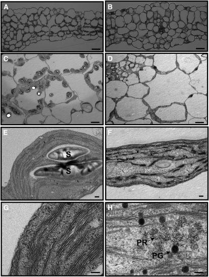
Figure 2.
Chloroplast Structures of the Wild Type and the pdtpi Mutant.
Panels show leaf and chloroplast structure of Columbia wild type ([A], [C], [E], and [G]) and pdtpi mutant ([B], [D], [F], and [H]). Plants were grown on MS plate for 5 d under continuous light and were then harvested for microscopy. Bars = 40 μ m in (A) and (B), 10 μ m in (C) and (D), 0.2 μ m in (E) and (F), and 100 nm in (G) and (H). Starch grains (S), plastoglobule (PG), and prolamellar body (PR) are indicated.
(A) and (B) Leaf structure observed under a light microscope (magnification × 500).
(C) to (H) Chloroplast ultrastructure from transmission electron microscopy.

